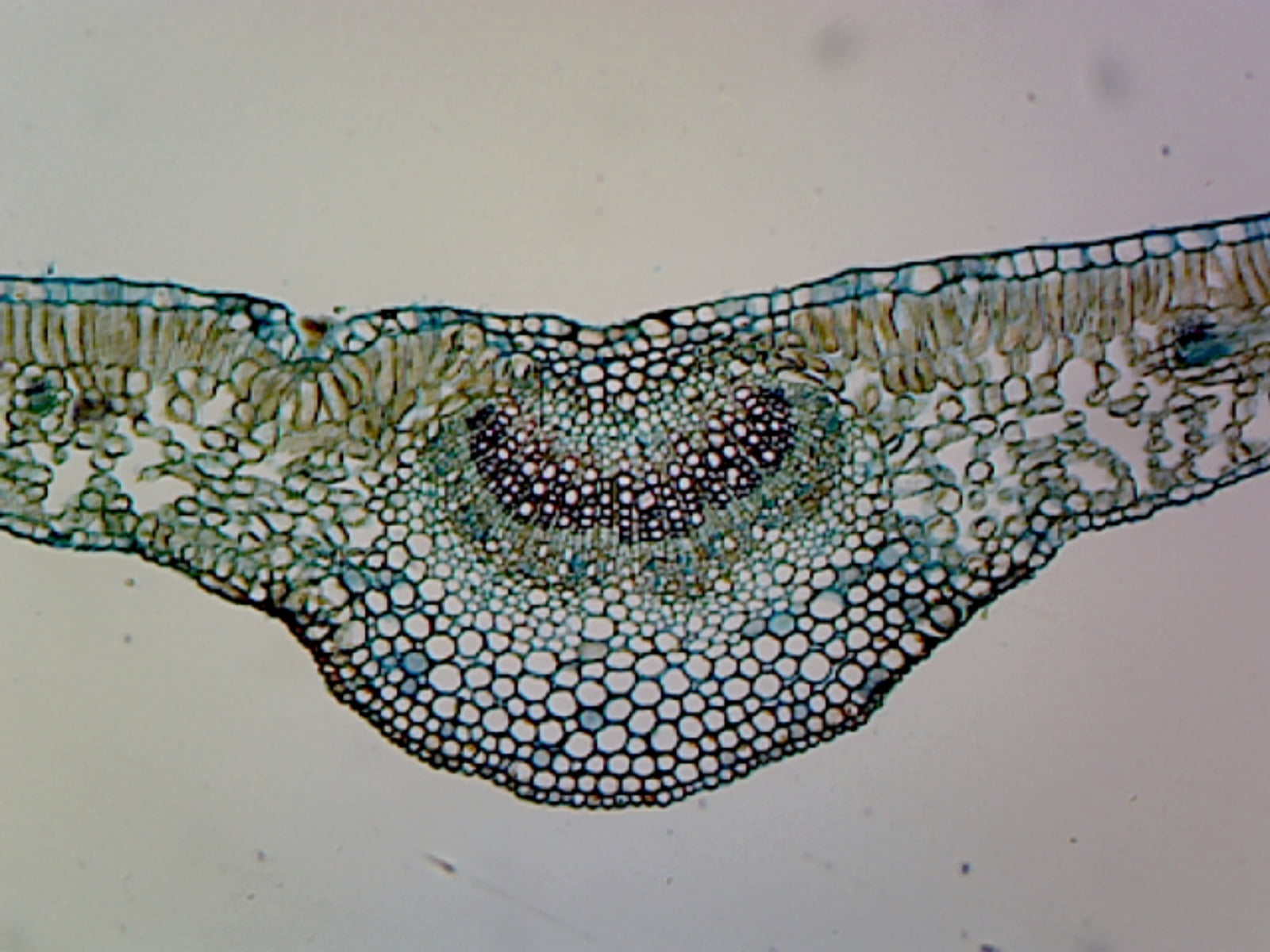
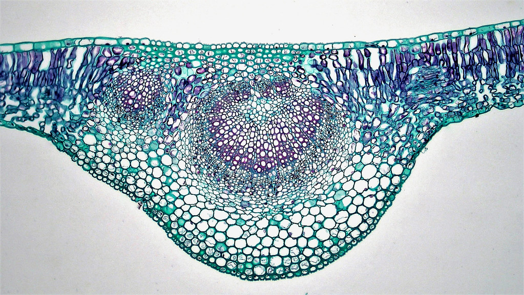
GSC International PS0079 Ligustrum Leaf; Showing Typical Mesophytic Dicot Leaf; Cross Section
Figure \(\PageIndex{15}\): Cross sections of a sun leaf (left) and shade leaf (right). The palisade parenchyma of the sun leaf consists of several layers, but there is only a single layer in the shade leaf. The chloroplasts (red dots) are also packed more densely in both the palisade and spongy mesophyll cells of the sun leaf compared to the.
Cross Section Of A Leaf Diagram Labeled Wiringopedia
A cross-section through a leaf Features of leaves and their functions The role of stomata The control gas exchange in the leaf. Each stoma can be open or closed, depending on how its guard.

Labeled cross section of a lilac leaf UWDC UWMadison Libraries
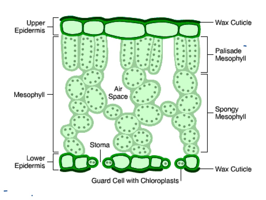
The leaves of dicotyledonous plants are arranged in a horizontal position, i.e., at a right angle to the rays of the sun so that they receive more light on the upper surface than the lower surface. The leaves with such an arrangement are known as dorsiventral or bifacial leaves.
:max_bytes(150000):strip_icc()/leaf_crossection-57bf24a83df78cc16e1f29fd.jpg)
Plant Leaves and Leaf Anatomy
The air space found between the spongy parenchyma cells allows gaseous exchange between the leaf and the outside atmosphere through the stomata. In aquatic plants, the intercellular spaces in the spongy parenchyma help the leaf float. Both layers of the mesophyll contain many chloroplasts. Figure 30.10. 1: Mesophyll: (a) (top) The central.
Leaf anatomy Royalty Free Vector Image VectorStock
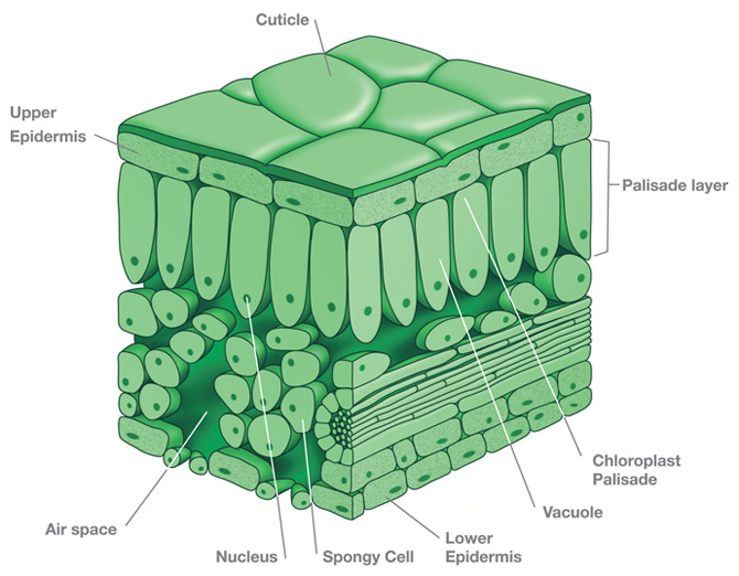
FIGURE 3. A cross section of a leaf. are responsible for most of the photosynthesis in the leaf and are called the palisade mesophyll. Located under the palisade mesophyll are loosely packed cells called the spongy mesophyll. The spongy mesophyll forms air spaces that hold raw materials to be used and products of
Cross section of a leaf blade of Nerium oleander a xeromorphic plant UWDC UWMadison
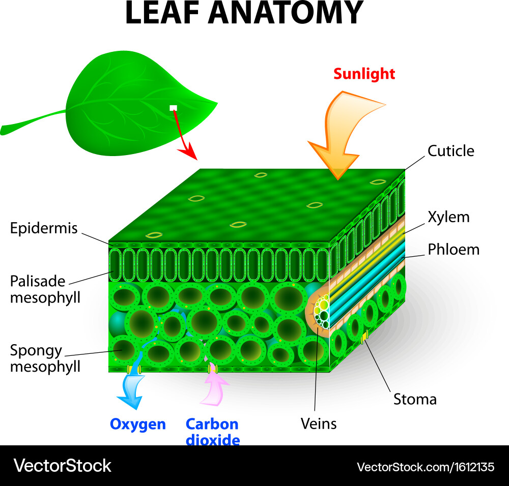
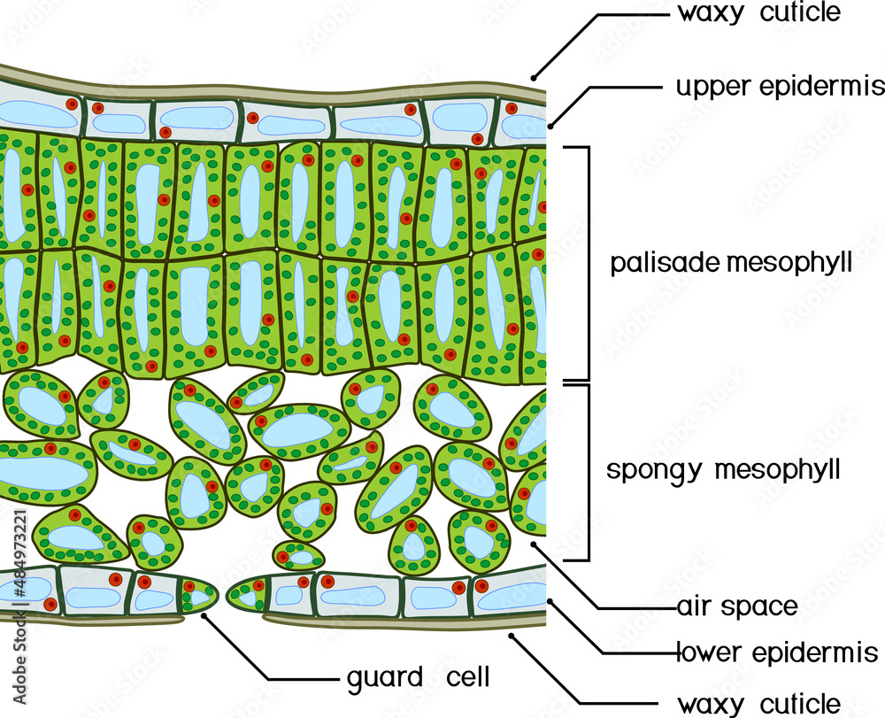
PHOTOSYNTHESIS [INTRO] Parts of a Leaf CrossSection of leaf.mov CROSS SECTION OF A LEAF Cuticle: A waxy layer that prevent water loss by evaporation. The cuticle is transparent and very thin to allow maximum light penetration. Upper Epidermis: A protective layer of cells that produces the cuticle.

Labeled Diagram Of A Leaf hubpages
Updated on November 04, 2019 Plant leaves help to sustain life on earth as they generate food for both plant and animal life. The leaf is the site of photosynthesis in plants. Photosynthesis is the process of absorbing energy from sunlight and using it to produce food in the form of sugars.

Leaf anatomical features (a) leaf cross section with dense crystal... Download Scientific Diagram
1 General characteristics 2 Morphology Toggle Morphology subsection 2.1 Basic leaf types 2.2 Arrangement on the stem 2.3 Divisions of the blade 2.4 Characteristics of the petiole 2.5 Veins 2.6 Morphology changes within a single plant 3 Anatomy Toggle Anatomy subsection 3.1 Medium-scale features 3.2 Small-scale features 3.3 Major leaf tissues

Cross Section Of A Dicot Leaf olddominiondesigningdivas
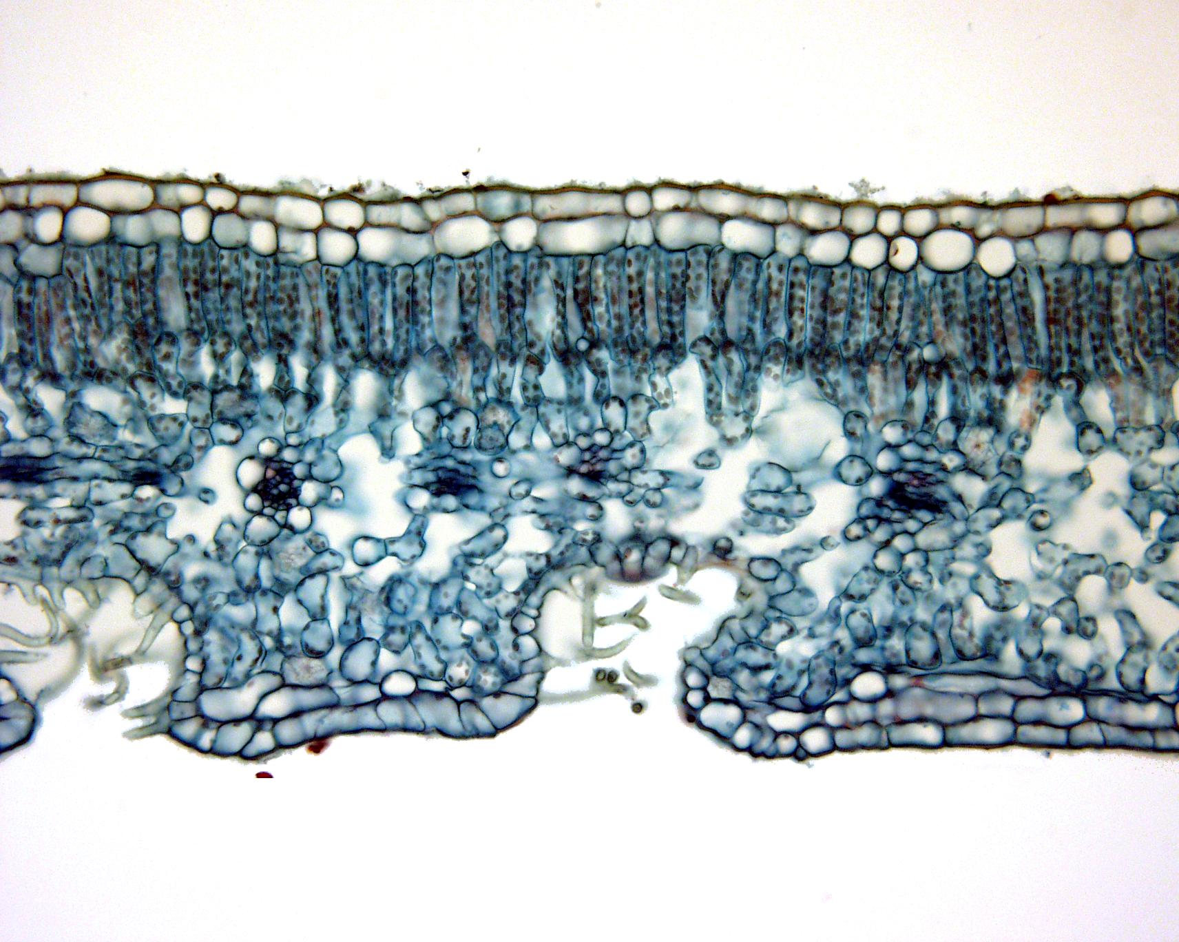
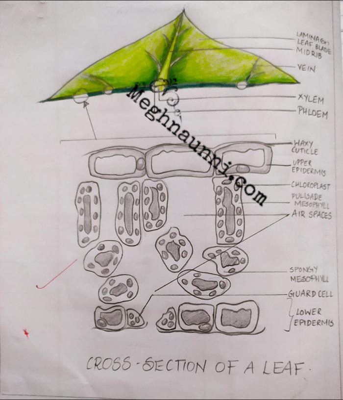
Cross-section of a leaf midvein. The section above was made through the midvein (large, central vein) of a lilac (Syringa) leaf. The midvein is in the center of the image, with xylem (water-conducting tissue) and phloem (food-conducting tissue) labelled. Surrounding the vascular tissues are thin-walled parenchyma cells and, under the upper and.
Plant Leaf Cross Section
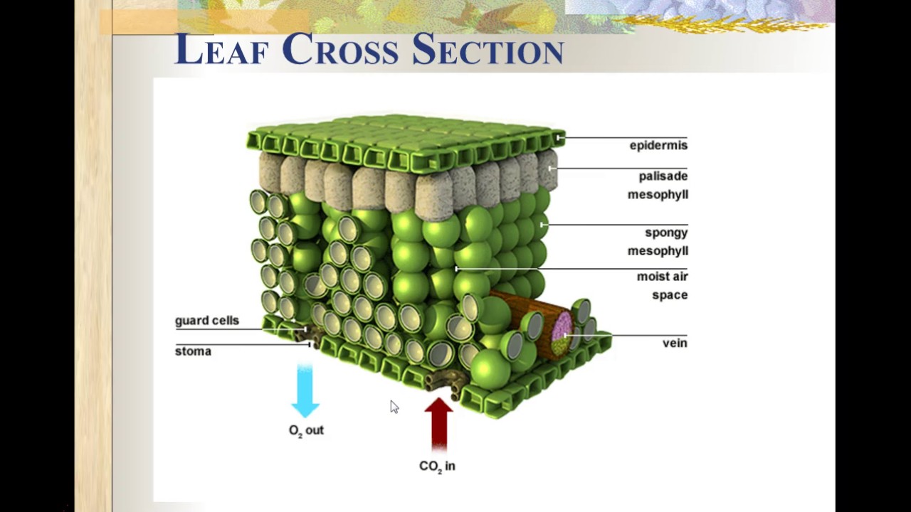
New version of this video: https://youtu.be/_y-HCi7mJjMThis is a description of a leaf cross section at the cellular level. Leaves contain a number of differ.
Plants Leaves
Leaf Structures Table. Diagram showing the cross-section of a leaf. The specialised cells in leaves have adaptive features which allow them to carry out a particular function in the plant; Adaptations of Plant Leaves for Photosynthesis Table. You've read 0 of your 0 free revision notes

what are the parts in a cross section of a leaf and what are their functions? Brainly.in
What are the primary components observed in a leaf cross section? What is the difference between palisade mesophyll and spongy mesophyll? How are stomata observed under the microscope? What is the role of the epidermal layer in a leaf? Why do leaves have air spaces in the spongy mesophyll?
Cross Section of a Leaf Biology Diagram
Figure 9.3. 2: Cross section of a hydrophytic leaf. Observe a prepared slide of a hydrophyte, such as Nymphaea, commonly called a water lily. Note the thin epidermal layer and the absence of stomata in the lower epidermis. In the spongy mesophyll, there are large pockets where air can be trapped.
37 leaf cross section diagram Diagram Online Source
Identify the parts of a typical leaf; Describe the internal structure and function of a leaf;. In this (c) light micrograph cross-section of an A. lyrata leaf, the guard cell pair is visible along with the large, sub-stomatal air space in the leaf. (credit: modification of work by Robert R. Wise; part c scale-bar data from Matt Russell).

Leaf CrossSection Crosssection through a dicot leaf, sho… Flickr
Figure \(\PageIndex{2}\): A cross section of a corn (Zea mays) leaf. See the caption in Fig. 13.2.3 for a detailed description of the features present. Photo by Maria Morrow, CC BY-NC. Figure \(\PageIndex{3}\): A cross section of a section of a corn leaf, labeled. The upper epidermis is composed of parenchyma cells that appear empty.
Dicotyledonous Leaf Cross Section designsbylima
(Cross Section in Above Right Photo) Color is produced by the balance of pigments in the leaf tissue and also by the distribution of pigments in the plastids as well as the air spaces inside of the leaf that scatter the light penetrating into the leaf.